Elevate Physical Theraphy & Fitness

physical therapy

Kink in your neck? Here’s what to do.

For the days when you wake up with a kinked neck because you slept funny, but still have to get to work – Here are a few at-home moves to try that could help you before you get in to see a PT!   Chin Tucks 1. Stand with your back against a wall 2. Move your chin towards your chest, while pushing the back of your head into the wall, and hold for 5 seconds as you feel a comfortable stretch from your neck to the base of your skull. Repeat 10 times.   Cervical Towel Traction 1) Lying on your back, grab the towel and place it behind your head so that the pull comes from the base of your skull. 2) Provide traction by scooting away from the door which puts tension through the rope and will give you a nice stretch through your neck. 🔹Start with 3-5 minutes. 🔹You can keep your legs bent or straighten them for more tension. 🔹When you are done, scoot back towards the door to decrease the tension and slowly take your head out of the towel traction. *Please see a PT at Elevate to get the proper set up for the towel traction device.   Sub-Occipital Stretch 1) Tuck your chin. 2) Place one hand on your chin and provide a gentle pressure to further tuck your chin. 3) Place your other hand on the back of your head and provide a gentle upward pull to provide a stretch at the base of your head.   3 Finger Cervical Flexion Rotation 1) Place your thumb on your sternum. Hold index, middle and ring fingers together. 2) Flex your chin down to your ring finger. Then rotate and look left. 3) Then ‘sweep’ down to fingers with chin, and rotate and look right. Stay in a pain free range of movement. As you progress, work your way to rotating at your index finger.   Upper Trapezius & Levator Scapulae Static Stretches Upper Trap: 1) Maintaining good sitting posture, side bend your head to one side, bringing your ear to your shoulder. 2) Take your arm and reach over and pull the opposite side of your head into further side bend Levator Scapulae: 1) Maintaining good sitting posture, rotate your head towards the angle of your armpit. 2) Take your arm and reach over to the base of your head and pull your head toward your armpit.

Is Your Athlete At Risk?

KNEE INJURIES – IS YOUR ATHLETE AT RISK?  THREE EASY TESTS! Movement Performance Assessments are a key measure to reveal young athletes’ risks for injury, learn what to do to bulletproof against them, and increase their odds of staying healthy and playing great during the season. PREVENTABLE KNEE INJURIES The knee is the most commonly injured joint in adolescent athletes.  Lost play time, not to mention pain, rehab, and potential long-term disability are common realities with knee injuries.  Some of the more common ones are patellofemoral pain (“runner’s knee”), IT Band Syndrome, and patellar tendinitis (“jumper’s knee”), ligament sprains, and meniscus tears.  Two additional injuries common in the skeletally immature are Osgood Schlatter’s disease and osteochondritis dessicans (OCD).  The less common (yet numbers are rising in our youth athletes) and more catastrophic injury is the ACL (anterior cruciate ligament) tear.    Seventy percent of ACL tears are non-contact injuries that occur during movements like cutting, twisting, decelerating, and landing from a jump, without making any contact with another player, in sports like soccer, basketball, lacrosse, football, volleyball, and tennis. It’s important to note too that there is a four to six-fold greater incidence in female athletes compared to males playing the same landing and cutting sports. Current research by many including Dr. Christopher Powers, Professor of Biokinesiology & Physical Therapy at USC, has revealed a number of modifiable risk factors associated with knee injuries, including the devastating ACL tear, i.e. weakness and poor control of the trunk, pelvis, hips, and knees, and poor body alignment during movement.  Dr. Timothy Hewett of The Mayo Clinic Orthopedic Biomechanics Lab describes these modifiable risk factors as: ligament dominance; quadriceps dominance; leg dominance; and trunk dominance, all of which can be changed to improve the odds of avoiding an injury.  These are preventable injuries. But how? TEST to uncover deficiencies associated with injury  Get key muscles strong Improve high-level balance  Train strength & balance with optimal movement  Incorporate into sport-specific movement (“neuromuscular control”) during warm-up and training  Good movers become better athletes and have fewer injuries.  TESTING There are numerous tests with complex scoring systems and criteria that we run for individuals and teams both in the clinic and on the field.  We use the latest evidence in testing and advanced technology to determine an athlete’s risk profile, we score the tests individually and look at them collectively to determine an athlete’s level of risk for injury.  Then we teach what to do.   If an athlete has been injured, we use these tests to determine return-to-sport readiness as well.   But here’s the good news… A  few simple tests can detect key risk factors associated with knee pain and injuries, including the devastating ACL tear. Step-Down Test 10 Second Tuck Jump Drop Jump Test The Step-Down Test is a low impact, basic indicator of an athlete’s single leg, dynamic strength, control, and preferred movement patterns.   What to look for that reveals greater risk for injury: Excessive leaning of the trunk (poor trunk control) Excessive drop of one side of the pelvis toward the ground (poor pelvic control) (#1 and #2 can often go hand-in-hand, as in the video, and even though the knee may not appear to drop inward (valgus angle), the combination of the two can create a valgus torque at the knee.) Rotating the thigh and the knee collapsing inward (dynamic valgus/poor hip utilization) Upright trunk (quadriceps dominance / poor hip utilization) The 10 Second Tuck Jump, formalized and studied by Dr. Gregory D. Myer, sports biomechanist at the Cincinnati Children’s Hospital Sports Medicine Biodynamics Center, is a ‘double leg ballistic/plyometric’ activity (jumping on both legs), increasing the demand to the system compared to the Step-Down Test.  It’s a great one to do on the field because it doesn’t require any equipment.  Have your athlete try it.  Take a video and use slow motion if possible. Jump for 10 seconds, trying to lift knees to hip height and trying to land on the same spot, without pausing. While watching the video, see if your athlete’s knees collapse inward (dynamic valgus), like the knees are kissing, as the feet hit or push off from the ground. If they do, like the young woman in the video, your athlete is at greater risk. If your athlete looks more like the young man in the video, the chances of injury are lower.   We actually score 10 criteria in this test, but here are five easy things to pick out that will give you insight into risk for injury: Knees collapsing inward / dynamic valgus Knees uneven and not reaching hip height Stiff knee landing Landing on one leg before the other Not remaining in the same ‘footprint’ throughout the 10 seconds The Drop Jump Test “before” video demonstrates “dynamic valgus” (inward collapse of the knees) in a volleyball athlete.  It’s one movement pattern associated with knee pain and injuries.  The “after” video demonstrates this same athlete after some strength, balance and movement training, using a band above the knees to cue and facilitate her gluteal muscles to control the movement and protect her knees. Dynamic valgus, combined with poor shock absorption and poor trunk control during cutting, twisting or landing can overload the ACL in a split second.  Wear and tear from dynamic valgus during daily activities and sports is also associated with the more common knee injuries mentioned.   COMPREHENSIVE MOVEMENT PERFORMANCE TESTING Try these three tests at home or on the field and see how you do.  Come in to do a comprehensive Movement Performance Assessment of 8 tests, including running and cutting sport-specific tests with video analysis with our PT team specializing in sports and biomechanics and learn what to do.  Taking time to learn which modifiable risk factors your athlete demonstrates and what to do about them will help your athlete avoid injury, achieve top performance, and STAY IN THE GAME.  

Hamstring Health, Part 2: Flexibility

Don’t “pull a hammy” Part 2! Hamstring Flexibility. Since a major key in injury prevention is flexibility, here are our favorite stretches that not only lengthen your hamstrings, but also protect your back while you’re doing them. 🔹Hamstring Hooklie Stretch 🔹Seated Hamstring Stretch 🔹Wall Assisted Hamstring Stretch 🔹Dynamic Walking Stretch   Hamstring Hooklie Stretch: While lying down on your back, hook a towel or strap under your foot and draw up your leg until a stretch is felt in the back of your leg. Keep both hips down on the floor and your knee slightly bent during the stretch. Hold for 60 seconds. Seated Hamstring Stretch – While seated, rest your heel on the floor with your knee straight and gently lean forward until a stretch is felt behind your knee/thigh. Keep your back straight! Hold for 60 seconds. Wall Assisted Hamstring Stretch – Place a leg up a wall while lying on your back. Your other leg should be positioned with a straight knee and resting on the floor through a doorway or hall. Make sure your hips stay even and on the floor. Hold for 60 seconds. Dynamic Walking Stretch – While keeping a flat back, step one foot in front of you with a straight leg and flexed foot. Hinge over at the hips and lean forward into your front leg until you feel a stretch. Hold for 3-5 seconds before switching to the other leg. Repeat 10 times on each leg.

Elevate’s Ankle Sprain Rehab

Sometimes it’s difficult to know if you’ve rolled your ankle or done something more that may require an x-ray. The Ottawa Ankle Rules help us decide whether or not an x-ray is warranted: Unable to put any weight on your injured side for 4 steps Tenderness at the base of your 5th toe. Tenderness 6 cm up from your ankle bone. If you have any of the above-mentioned rules, then you should get an x-ray to rule out a fracture. Lateral Ankle Sprains can vary in their severity. Clinically, ankle sprains  are generally classified into three groups: Grade 1: A stretch of the ligament that does not result in a tear. There is minimal swelling and tenderness, mild or no loss of function, and no mechanical instability of the ankle. Generally associated with strain to the ATFL. Grade 2: A partial tear of the ligament with moderate pain, tenderness, and swelling. There is some loss of function and presence of mild to moderate mechanical instability. Generally associated with strain to both the ATFL and CFL. Grade 3: A complete tear of the ligament with significant bruising, swelling, and tenderness. There may be an inability to bear weight on that foot, and there is significant instability of the ankle. Generally associated with strain to the ATFL, CFL, and potentially the PTFL as well. There may be swelling, bruising, and tenderness in your foot and ankle. It is important to note that the amount of swelling and bruising is not always indicative of severity of tissue damage. The best time to have a physical examination by your Physical Therapist is 4-5 days after your injury.     After an ankle sprain it is important to work on ankle mobility, motor control, and balance. The following exercises show a few examples of some exercises used to address those impairments. 1.Ankle CARs (Controlled Articular Rotations) – helps to regain mobility and useful as an assessment of motor control 2.CRAs Correctives – if you find an area of movement during your CARs that is more challenging or in uncontrolled (might be shaky or not a smooth path of movement), then you can work in that uncontrolled range to help improve your control over that particular range 3.Single Leg Balance – the video shows progressively more challenging ways of working on single leg balance. If you are in a lot of pain still and cannot bear your full weight on your injured side, you can do these exercises on your other side since there is a known cross-over effect for these exercises (training one side of the body will also have a training effect on the other side of the body) 4.Multi-directional reach – this works on your single leg stability in multiple planes of movement   In order to promote healing and help reduce the risk of future injury, it is important to progressively overload the tissues that support the ankle in order to create adaptation. The exercises in this video demonstrate a few ways that you can target loading of ankle inversion. Medial/Lateral Lunge Three Way Step Down Cross-Over Step Up Single Leg Medial/Lateral Reaches Single Leg RDL Single Leg Airplane With Rotations Single Leg Golfer RDL’s Single Leg Lateral Med Ball Toss You can gradually add weight to these to make them more challenging.

Concussions 101: What You Should Know – A Four-Part Series

Concussions have become a highly discussed and debated topic. Because of our increased awareness, the diagnosis of concussion has increased dramatically at all levels of athletics and in the general population. However, many athletes, parents, and even clinicians lack a good understanding of the underlying condition, the several types of concussion, and the appropriate ways to properly evaluate and manage the condition. WHAT IS A CONCUSSION? A concussion is a functional injury of the brain that results in altered metabolic activity within the brain. This alteration involves changes in chemical activity, increased energy demand, and decreased blood flow within the brain. Simply put, the combination of increased energy demand and decreased blood flow results in an “energy crisis” that impairs optimal function and leads to the symptoms of a concussion. Many of these changes occur within the first 24 hours of the injury, but some of these changes can persist for multiple weeks! Contrary to widespread belief, a concussion does not involve any structural damage to the brain or surrounding structures of the head and neck. Other injuries to the head and neck may occur in conjunction with a concussion but are not required to be diagnosed with a concussion, which is diagnosed based on the individual’s signs and symptoms. Therefore, if a concussion is suspected it is imperative to be evaluated by a medical professional that can guide appropriate care and rule out the possibility of a structural injury that may require diagnostic imaging. However, if a head or neck injury is not suspected, then diagnostic imaging is not indicated, and diagnosis is based on a thorough clinical examination. WHAT CAUSES A CONCUSSION? Concussions can occur through a variety of mechanisms. Although most common in impact sports, they can also occur in non-impact sports and in incidents such as motor vehicle accidents. Typically, a concussion occurs when an impact is made with the head, or the head and neck undergo a violent acceleration or deceleration motion. These motions can place stresses on the structures within the brain that result in injury and responds with altered metabolic activity. It is important to remember that a concussion does not always require a violent motion or loss of consciousness. Even seemingly innocent injuries can lead to a concussion! Furthermore, signs and symptoms do not always present immediately, so it is important to be aware of and monitor for the signs and symptoms and seek medical care if suspected. WHAT ARE THE SIGNS AND SYMPTOMS OF A CONCUSSION? Typical signs and symptoms of a concussion can vary between individuals. The most common symptom of a concussion is a headache, but other common symptoms may include: Additionally, symptom presentation and recovery can vary drastically between individuals and may be based on age, gender, activity level, and history of prior injury, among other factors. For instance, symptoms may be exaggerated in younger individuals due to an immature neurologic system. On average, professional athletes become asymptomatic in 1-2 days, collegiate athletes in 2-7 days, and high school athletes in 1-2 weeks! And, while most people recover in less than 2 weeks, approximately 20% of individuals take more than 3 weeks to recover! This is referred to as post-concussion syndrome and can lead to persistent symptoms that not only impact return to athletic activities but can impair mental performance and cause persistent pain. In these scenarios it is especially important to seek medical advice from a qualified medical provider that can accurately identify the type of concussion, contributing factors, and appropriate management options. WHAT TO DO… Now that you are more informed about the potential causative factors and signs and symptoms related to concussion, it is important to recognize when a concussion might be present and be evaluated by a medical professional if it is suspected. If sustained at a school-related sporting event this may be performed by the Athletic Trainer, but, if one is not present, a physician that specializes in concussion is a great option. They can decide if further testing is warranted and refer to Physical Therapy! As we will soon see in the next segment in this four-part series, a concussion is not as simple as we once thought, and Physical Therapy can be a fantastic treatment option to promote return to function.

Fruit: Nature’s Answer to Your Snack Cravings

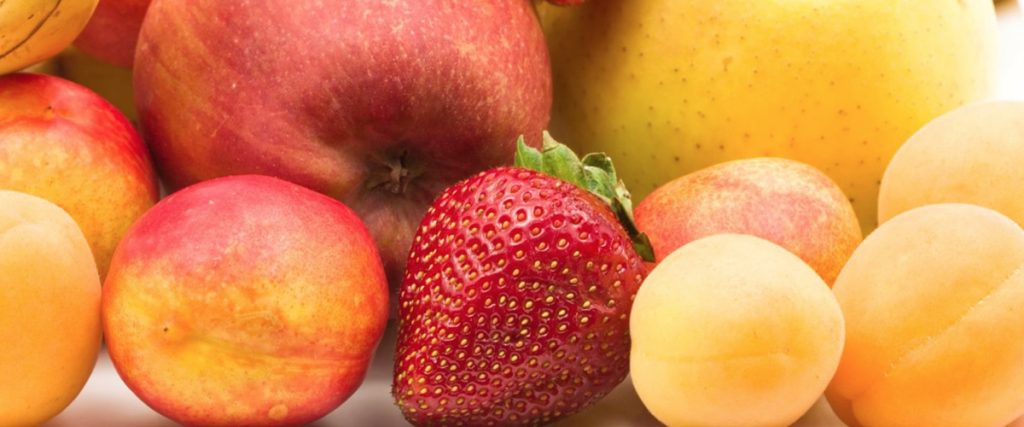
By Kyle Blix, CPT, Cert. Ace Fitness Nutrition Specialist Are you working hard in the gym but not seeing the changes you’re going for with your weight or body composition? It may very well be your diet–and more specifically, your snacks. Perhaps the greatest contributor to seeing results in with any exercise program is the diet. A week of hard lifting and strenuous cardio can be seriously undercut by poor food decisions. This is because food plays such a critical role in rebuilding the cells of the human body. As a personal trainer and nutrition counselor, I stress the importance of eating real, whole, unprocessed foods as much as possible, because they’re proven to be the most nutritionally dense and thus keep the body healthy and lean.(1) When strategizing for proper nutrition, one of the most helpful questions to ask first is:  What are the foods that you know, without a doubt, are holding you back? And more specifically, what are the snack foods that you’re eating in-between and after your meals? Often times people will get into a whole food diet only to go off the rails when it comes to their snack habits, which in turn can sabotage their progress of sculpting a lean physique. Here,  we turn to nature for the solution–fruit.  When used as a snack replacement, fruit is one of the easiest ways to keep your body healthy and lean without sacrificing flavor and satisfaction. Fruits are nature’s ultimate snack hack. To better understand just how fruit interacts with our bodies, we must address fructose. Simply put, fructose is fruit sugar. It’s a monosaccharide, the simplest form of carbohydrate, and thus it digests very easily.(²)  Fructose is also found in processed foods as well, more commonly under the name high fructose corn syrup (HFCS). It’s a cheap way to sweeten processed foods and is found in a variety of products. However, there’s a big difference between HFCS and natural fructose from fruit.  The negative effects of high fructose in the diet are known to cause metabolic issues, such as obesity, high blood pressure and type 2 diabetes. But the naturally occurring fructose in fruit is completely different. Unlike HFCS, fructose does not cause a rapid rise and subsequent drop in blood glucose levels, giving it a fairly low glycemic load. Glycemic load is the measure of how a carbohydrate impacts your blood glucose levels.(³)  Fruit has a fairly low glycemic load and contains a good amount fiber and water, which, when consumed together, help to mitigate the effects of fructose on blood sugar. For this reason, most fruits take a while to digest and hit the liver-insulin system slowly.(4)  This is beneficial because consuming foods with a low glycemic load reduce the chance of health problems like diabetes, hypertension, obesity and heart disease. Compare eating two apples, with a total of 38g of fructose sugar, to a can of Coke containing 39g of HFCS or cane sugar. One is going to keep you satisfied and fuller, and the other is going to cause a huge spike and crash. They both have completely different effects on the body. The nutrient density of fruits cannot be ignored. They’re rich in fiber, vitamins, minerals, as well as a plethora of phytonutrients and antioxidants. Plus they’re incredibly filling, and delicious. Fruits have been shown in multiple studies to reduce the risk for type 2 diabetes, heart-disease and as well as different types of cancer. In one particular study, research found that the risk of heart disease is reduced by 7% for each daily portion of fruit.(5) Fiber especially has many benefits. The soluble fiber found in fruits has been shown in several studies to reduce cholesterol levels as well as slow down the absorption of carbohydrates.(6)  This benefit carries over into weight loss as well, as fiber plays a critical role in increasing satiety, which in turn leads to consuming fewer calories. On the caloric front, fruits come with more good news! They’re relatively low in calories, which means you can consume a variety of fruits before coming anywhere close to what the average bag of chips would cost you, for example. The average apple comes in at 95 calories, a banana 100 calories, an orange 45 calories and a carton of strawberries at 145 calories. When you habitually eat fruit, you’ll be less inclined to turn to snacks that are devoid of nutritional value. To drive it home, the most useful way to work fruits into your diet is to use them as a complete snack replacement.  Fruits are nature’s ultimate snack-hack. Take any and all sweet snacks that you would normally eat after a lunch or dinner and simply replace them with a variety of fruits. For example, you could pack two bananas and a Tupperware of berries to take with you to work. Or toss them in a smoothie along with a plant-based protein powder for a meal replacement. They’re portable, tasty, and most importantly, incredibly nutrient-dense. You could even start with eating only fruit for breakfast and you would already be on your way to a much healthier day than if you picked a sugary cereal or a bagel. Armed with this knowledge, try incorporating more fruit into your diet combined with your exercise program to see those results you’re hoping for and realize the multitude of health benefits from fruit!   References: 1: Slavin, Joanne L., and Beate Lloyd. “Health Benefits of Fruits and Vegetables.” Advances in Nutrition 3.4 (2012): 506–516. PMC. Web. 16 Mar. 2018. 2: The Britannica – https://www.britannica.com/science/monosaccharide 3:Eleazu, Chinedum Ogbonnaya. “The Concept of Low Glycemic Index and Glycemic Load Foods as Panacea for Type 2 Diabetes Mellitus; Prospects, Challenges and Solutions.” African Health Sciences 16.2 (2016): 468–479. PMC. Web. 16 Mar. 2018. 4:Tappy L, Lê KA. Metabolic effects of fructose and the worldwide increase in obesity. Physiol Rev. 2010 Jan;90(1):23-46. doi: 10.1152/physrev.00019.2009. Review. PubMed PMID: 20086073. 5: Luc Dauchet, Philippe Amouyel, Serge Hercberg, Jean Dallongeville; Fruit and Vegetable Consumption and Risk of Coronary

What is Achilles Tendinopathy?

Achilles Tendinopathy is a common injury in runners and other athletes. It is generally classified as localized pain in the tendon and/or heel that is worse with increased loads through the tendon (a heel raise hurts more than standing, hopping feels worse than heel raises). People also usually report their symptoms are worse in the morning during the first few steps right after getting out of bed. Although this condition has historically been known as a “tendinitis”, it is now being referred to as “tendinopathy”. The suffix “itis” means “inflammation” and in this condition, there are generally no inflammatory cells present and causing the symptoms. Rather, it is more of a reactive process to overload. Tendinopathy refers to any problem within the tendon and its cells, and is therefore a more fitting name than tendinitis. Insertional vs Mid-Portion Achilles Tendinopathy There are two different forms of Achilles Tendinopathy and it is important to know the difference between the two of them because some of the exercises vary slightly for each. In Insertional Achilles Tendinopathy, there is generally pain where the Achilles Tendon inserts on the calcaneus (the heel bone). Performing a calf stretch, walking barefoot, squatting, and uphill walking may all be aggravating factors. In Mid-Portion Achilles Tendinopathy, the pain is generally located 2-6 cm above the heel. Aggravating factors may include running, heel raises, and hopping. If you think you may have Achilles Tendinopathy, it is important to have a thorough evaluation by a Physical Therapist so that they can rule out other potential causes of your pain, and help design an individualized rehabilitation program for you based on the Stage of Tendinopathy you are currently presenting in, what your personal goals are, what type of Achilles Tendinopathy you have, and any other biomechanical or movement issues that need to be addressed. What causes Achilles Tendinopathy? The single greatest cause Achilles Tendinopathy is training errors. Sudden increases in activity, mileage, speed, incline running, changing stride or cadence can all lead to tendinopathy. Remember, your body is always in a balancing act of load/wear and rest/repair. Increasing training load or activity too much without enough rest can tip your tendons too far towards the load/wear side of the spectrum and can cause tendinopathy. A good, general rule of thumb to follow is to not exceed increases in training load by more than 10% each week. That means that if you have been able to comfortably run 20 miles per week for the past month, then you can increase your next week’s mileage to 22 miles without increasing your risk of injury. Of course, each person is different and it is therefore advisable to track your training load with the help of a Doctor of Physical Therapy or Personal Trainer to make sure you are progressing in a safe and gradual manner. Some other risk factors for Achilles Tendinopathy include a previous history of Achilles Tendinopathy, recent injury, age, gender, muscle power/strength, poor dorsiflexion, excessive pronation, and weight gain. Another risk factor that often goes unmentioned is taking antibiotics from the Fluoroquinolone family. These medications are used to treat a range of illnesses including respiratory infections and urinary tract infections. Some of these medications commonly prescribed are Ciprofloxacin (Cipro), Gemifloxacin (Factive), Ofloxacin (Floxin), Moxifloxacin (Avelox), Levofloxacin (Levaquin), and Norfloxacin (Noroxin). This risk increases with higher dosages of the medication. When compared to other classes of antibiotics, fluoroquinolones demonstrate a 3.8-fold greater risk of developing Achilles tendinopathy/rupture. Patients treated with fluoroquinolones have a 1.3-fold increased risk of tendon rupture compared to those not taking them, and that risk increases to a 46-fold greater predisposition if patients are also exposed to corticosteroids simultaneously. Age greater than 60 also seems to increase the risk of tendon rupture with fluoroquinolone use. If you have recently taken, or are currently taking any of these medications, ask your Physician or Pharmacist for information about the increased risk of tendon ruptures while on these medications. It is also important that you tell your Physical Therapist and Personal Trainer if you are on (or have recently been on) any of these medications so that the necessary load modifications can be made. The body is constantly in an equilibrium between load (or use)/wear and rest/repair. Whenever you perform a bicep curl, for example, you are applying load to the bicep muscle and tendon. Throughout the performance of the curls, the bicep is subjected to loads which cause micro tears within the muscle belly. When you are done working out, and give your bicep time to recover, those micro tears begin to repair and then become adapted to the loads you have just subjected them to. The key here is that you must give your body ample time to recover between bouts of loading. The same is true in all bodily tissues. When you apply a given load, there is a period of wear and then the tissue needs time to recover and repair during which time it becomes adapted to handle the loads you applied to it. When the scale begins to tip too heavily towards the load/wear side without enough rest/repair, the tissues you loaded are at an increased risk of injury. In tendinopathy, when tendon loading exceeds the tendon’s capacity (you ask it to do more than it is able to at that particular point in time), without sufficient rest, it develops into a tendinopathic tendon which then becomes less capable of handling load. The Stages of Tendinopathy: Before delving into how to treat Achilles Tendinopathy, it is important to understand the different stages of tendinopathy. These stages are classified differently, but they are really more of a continuum with load being the main driver along the spectrum. The Reactive Stage: The Reactive Stage of Tendinopathy is the tendon’s immediate response to overload. It can be painful and irritable in this phase. This is something that many people have experienced. If you have ever spent a day walking around Disney Land and then noticed by the end of

Congrats to Elevate Runner Claudia Lane!

Claudia Lane recently won first place in the Foot Locker Cross Country National Championship, completing a 5k run in only 17:04.8! Congratulations on an amazing finish, from the entire Elevate team! More photos from the race and all of the finishing times are posted on the Foot Locker National Championship website.

Rethinking Movement: Why You Should See A Physical Therapist Every Year

Why is it that superbly fit athletes can find themselves in as much back, knee, or neck pain as their flabby fans, who sit at desks all day long then watch sports from overstuffed sofas? “When you do an activity over and over again, your body adapts to that activity,” warns Dr. Shirley Sahrmann, professor emerita of physical therapy at Washington University School of Medicine. “If you play tennis, your arm gets bigger on that side; if you do karate you get adaptations in your hip and leg. Even if you just sit, you lean, you slump, your neck goes forward.” Either your body fails to build up musculature to support itself, or it overbuilds certain muscles and throws off the symmetry your skeleton craves. That’s why Sahrmann wants to see an annual physical therapy exam become as routine as a dental checkup. “We go to the dentist twice a year and spend thousands to straighten our teeth, and all we do with them is eat and talk. Meanwhile the rest of our body’s just hanging out there.” People think of PT as something generic their doctor orders after an injury, she says. But by analyzing the way you walk, bend, sit, and carry yourself, physical therapists can prevent injuries and head off future surgeries and chronic pain. “Kids don’t sit correctly, they slump, so they wind up sitting on the middle of their back,” she says. “We have these little bones on our bottom where we are supposed to sit and keep our spine erect. When you slump, the muscles get stretched out, and they’re not going to function optimally.” A temporary phase? Maybe. But “bones adapt to the alignment that you keep them in,” Sahrmann points out, “and your spine becomes shaped like that.” Watching teenagers walk makes her crazy: “They are not using what we consider a normal gait. They walk without bending at the hip and knee and pushing off. They shuffle. And they sway back—their shoulders are behind their hips—so their gluteal muscles don’t work as much as they should. All of these little cultural changes in sitting posture, what’s considered cool—even the clothing.” For a while there, she says, “the new waistline was the gluteal fold! And how do you walk when your legs are strapped together with a belt? Their knees get caught in the crotch of their pants—it’s hysterical. But it’s also not good.” In years past, there was little appreciation of how lifestyle affected your health. “My family thought they just got diabetes or hypertension; it had nothing to do with the cans of Crisco my grandmother cooked with.” The way we move and align ourselves is just as important as what we eat, she says. “There is complexity to movement, and you can do it right or wrong.” I bring up ergonomics—surely that’s helped? “It’s not just whether the setup is right,” she points out. “It’s what you bring to that setup and what you do when you’re not there.” We’re designed to keep changing position, not sit frozen in the perfect chair. Even working out requires real knowledge, if you want to lift weights or do aerobic training without compromising the performance of all your other joints. Sahrmann’s one of the nation’s pioneers in pushing the concept of a movement system, emphasizing the subtle, necessary interconnections of muscles and bones and nerves but also heart, lungs, and the endocrine system. Her career has spanned more than half a century, and its twists and turns led her to see the body whole. She began work at the end of the polio era and spent nine years taking care of patients who’d suffered head injuries or strokes. In order to understand the disordered motor control of patients with central nervous system lesions, she left clinical practice to earn a Ph.D. in neurobiology. Then serendipity sent her a different kind of patient: athletes who had musculoskeletal pain.  “I started teaching them to move differently, and they got better for reasons I didn’t understand,” she says. “I’ve spent a good many years with my colleagues at Wash.U. working to analyze these relationships between movement and musculoskeletal pain.” She’s written two books and talked herself hoarse, urging people to see the body’s movement as systemic. Now that approach is finally catching hold, not just here but nationally and internationally. But she’ll know she’s really succeeded when PT evaluations are annual, and there are formal diagnoses based on movement patterns that consistently cause pain: flexion syndrome, when the back bows out; extension syndrome when it bows in and hunches you over; tibiofemoral rotation that can lead to knee problems. “We all move differently,” she says. “I’ve seen patients whose feet are so callused I don’t know how they put their shoes on, and I’ve seen marathon runners with no calluses at all. “You need to have an exam by a body expert at least once a year,” she finishes crisply. “Even if insurance doesn’t cover it, the cost is no more than you’d pay a personal trainer. I think we could substantially reduce the number of injuries and slow the process of osteoarthritis as people age.” She’s not saying arthritis can be eliminated altogether—but it can be delayed and its effects minimized. “There’s evidence that if a joint is lax, or you have injury, or your muscles are weak, you can get these arthritic changes.” Move right, and you lower the chance of injury—whether you’re an Olympian or a couch potato. By Jeannette Cooperman May 11, 2015 / St. Louis Magazine  

Parent Confessions… I Hurt My Little Leaguer’s Arm

Little League baseball season is in full ‘swing’. Look out for overuse injuries in your child, and check this out from littleleague.org of one parent’s account of his child’s overuse and need for Tommy John surgery… he thought it could never happen to his child. If your baseball player is experiencing elbow pain, come in for a PT evaluation, see your pediatrician, orthopedist, or sports medicine doctor. We’re reaching epidemic proportions in the number of youth players requiring this surgery. Be aware, and prevent it from happening to your child. __________________________________________ Parent Confessions… I Hurt My Little Leaguer’s Arm I remember the day my wife gave me an article about kids having Tommy John surgery. I read it, and kind of blew it off. Not my kid I thought. He’ll just take it easy once in a while. He’ll be fine. Well, it ended up being my kid. All that talent… gone. I blamed others. My wife blamed me. She was right. As an 11-year-old, my son didn’t lose a game on the mound. He pitched two no-hitters. Even the high-school coach came to see him pitch. “Lots of talent, AND he’s a lefty!” he said after a game. I kept picturing my son sitting at a table, signing a Division 1 Letter of Intent. We started practicing for his final year of Little League the very next day after his last game as an 11-year-old. I convinced him basketball was taking up too much time, and that if he concentrated only on baseball, he might get a free ride somewhere. I know now that was a mistake. Throughout the fall and winter, he worked with a pitching coach, and he continued with his travel ball team. We live in North Carolina, so we play pretty much year round. He was the ace on the travel ball team. The manager pitched him… A LOT. My son won… A LOT. We concluded a travel ball tournament on a Friday night. He pitched five innings. Got the W. The next day was “Opening Day” for Little League. My son was so excited. His coach came up to me before the game, and asked if my son had been pitching a lot on his travel ball team. “Nah,” I said. He kind of just looked at me. “I don’t want to risk injuring his arm,” he said. “You need to tell me if he’s pitching too much on the other team.” “He’s fine,” I told him. “Opening day, Coach. Gotta go with your number one, right?” He did. We won. Me and my son’s Little League coach had the same type of conversation one more time during the year. He had those talks with my son, too. My son, like me, said his arm was fine. Like most kids, he wasn’t going to say if he felt something a little weird in his elbow. My son was 3-0. He was scheduled to start against the best team in the league. He was excited. I was excited, until that is, another kid took the mound in his place. I poked my head in the dugout. “I’m shutting him down,” said the manager. I asked why. “His arm is dead! I told you to tell if he’s pitching a lot with other teams!” I ended up resting him a little. That season, he only pitched one more time for his Little League team. I rested him some more. He played 50/70, and continued travel ball. His travel ball coach pitched him. I shouldn’t have let him. His arm felt good one day, tired the next. We rested, iced it, and heated it. Sometimes he threw like his old self. Most times, he didn’t. At 14, we went to the doctor. And at only 15, my son had Tommy John. He will never be the same, and that Letter of Intent that I was so intent on will never come.