Elevate Physical Theraphy & Fitness

youth

Is Your Athlete At Risk?

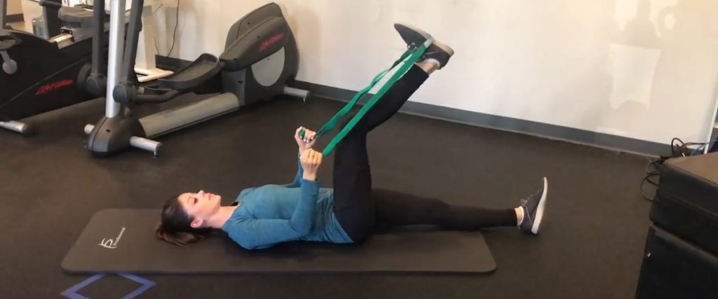
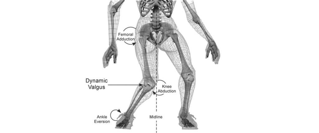
KNEE INJURIES – IS YOUR ATHLETE AT RISK?  THREE EASY TESTS! Movement Performance Assessments are a key measure to reveal young athletes’ risks for injury, learn what to do to bulletproof against them, and increase their odds of staying healthy and playing great during the season. PREVENTABLE KNEE INJURIES The knee is the most commonly injured joint in adolescent athletes.  Lost play time, not to mention pain, rehab, and potential long-term disability are common realities with knee injuries.  Some of the more common ones are patellofemoral pain (“runner’s knee”), IT Band Syndrome, and patellar tendinitis (“jumper’s knee”), ligament sprains, and meniscus tears.  Two additional injuries common in the skeletally immature are Osgood Schlatter’s disease and osteochondritis dessicans (OCD).  The less common (yet numbers are rising in our youth athletes) and more catastrophic injury is the ACL (anterior cruciate ligament) tear.    Seventy percent of ACL tears are non-contact injuries that occur during movements like cutting, twisting, decelerating, and landing from a jump, without making any contact with another player, in sports like soccer, basketball, lacrosse, football, volleyball, and tennis. It’s important to note too that there is a four to six-fold greater incidence in female athletes compared to males playing the same landing and cutting sports. Current research by many including Dr. Christopher Powers, Professor of Biokinesiology & Physical Therapy at USC, has revealed a number of modifiable risk factors associated with knee injuries, including the devastating ACL tear, i.e. weakness and poor control of the trunk, pelvis, hips, and knees, and poor body alignment during movement.  Dr. Timothy Hewett of The Mayo Clinic Orthopedic Biomechanics Lab describes these modifiable risk factors as: ligament dominance; quadriceps dominance; leg dominance; and trunk dominance, all of which can be changed to improve the odds of avoiding an injury.  These are preventable injuries. But how? TEST to uncover deficiencies associated with injury  Get key muscles strong Improve high-level balance  Train strength & balance with optimal movement  Incorporate into sport-specific movement (“neuromuscular control”) during warm-up and training  Good movers become better athletes and have fewer injuries.  TESTING There are numerous tests with complex scoring systems and criteria that we run for individuals and teams both in the clinic and on the field.  We use the latest evidence in testing and advanced technology to determine an athlete’s risk profile, we score the tests individually and look at them collectively to determine an athlete’s level of risk for injury.  Then we teach what to do.   If an athlete has been injured, we use these tests to determine return-to-sport readiness as well.   But here’s the good news… A  few simple tests can detect key risk factors associated with knee pain and injuries, including the devastating ACL tear. Step-Down Test 10 Second Tuck Jump Drop Jump Test The Step-Down Test is a low impact, basic indicator of an athlete’s single leg, dynamic strength, control, and preferred movement patterns.   What to look for that reveals greater risk for injury: Excessive leaning of the trunk (poor trunk control) Excessive drop of one side of the pelvis toward the ground (poor pelvic control) (#1 and #2 can often go hand-in-hand, as in the video, and even though the knee may not appear to drop inward (valgus angle), the combination of the two can create a valgus torque at the knee.) Rotating the thigh and the knee collapsing inward (dynamic valgus/poor hip utilization) Upright trunk (quadriceps dominance / poor hip utilization) The 10 Second Tuck Jump, formalized and studied by Dr. Gregory D. Myer, sports biomechanist at the Cincinnati Children’s Hospital Sports Medicine Biodynamics Center, is a ‘double leg ballistic/plyometric’ activity (jumping on both legs), increasing the demand to the system compared to the Step-Down Test.  It’s a great one to do on the field because it doesn’t require any equipment.  Have your athlete try it.  Take a video and use slow motion if possible. Jump for 10 seconds, trying to lift knees to hip height and trying to land on the same spot, without pausing. While watching the video, see if your athlete’s knees collapse inward (dynamic valgus), like the knees are kissing, as the feet hit or push off from the ground. If they do, like the young woman in the video, your athlete is at greater risk. If your athlete looks more like the young man in the video, the chances of injury are lower.   We actually score 10 criteria in this test, but here are five easy things to pick out that will give you insight into risk for injury: Knees collapsing inward / dynamic valgus Knees uneven and not reaching hip height Stiff knee landing Landing on one leg before the other Not remaining in the same ‘footprint’ throughout the 10 seconds The Drop Jump Test “before” video demonstrates “dynamic valgus” (inward collapse of the knees) in a volleyball athlete.  It’s one movement pattern associated with knee pain and injuries.  The “after” video demonstrates this same athlete after some strength, balance and movement training, using a band above the knees to cue and facilitate her gluteal muscles to control the movement and protect her knees. Dynamic valgus, combined with poor shock absorption and poor trunk control during cutting, twisting or landing can overload the ACL in a split second.  Wear and tear from dynamic valgus during daily activities and sports is also associated with the more common knee injuries mentioned.   COMPREHENSIVE MOVEMENT PERFORMANCE TESTING Try these three tests at home or on the field and see how you do.  Come in to do a comprehensive Movement Performance Assessment of 8 tests, including running and cutting sport-specific tests with video analysis with our PT team specializing in sports and biomechanics and learn what to do.  Taking time to learn which modifiable risk factors your athlete demonstrates and what to do about them will help your athlete avoid injury, achieve top performance, and STAY IN THE GAME.  

Hamstring Health, Part 2: Flexibility

Don’t “pull a hammy” Part 2! Hamstring Flexibility. Since a major key in injury prevention is flexibility, here are our favorite stretches that not only lengthen your hamstrings, but also protect your back while you’re doing them. 🔹Hamstring Hooklie Stretch 🔹Seated Hamstring Stretch 🔹Wall Assisted Hamstring Stretch 🔹Dynamic Walking Stretch   Hamstring Hooklie Stretch: While lying down on your back, hook a towel or strap under your foot and draw up your leg until a stretch is felt in the back of your leg. Keep both hips down on the floor and your knee slightly bent during the stretch. Hold for 60 seconds. Seated Hamstring Stretch – While seated, rest your heel on the floor with your knee straight and gently lean forward until a stretch is felt behind your knee/thigh. Keep your back straight! Hold for 60 seconds. Wall Assisted Hamstring Stretch – Place a leg up a wall while lying on your back. Your other leg should be positioned with a straight knee and resting on the floor through a doorway or hall. Make sure your hips stay even and on the floor. Hold for 60 seconds. Dynamic Walking Stretch – While keeping a flat back, step one foot in front of you with a straight leg and flexed foot. Hinge over at the hips and lean forward into your front leg until you feel a stretch. Hold for 3-5 seconds before switching to the other leg. Repeat 10 times on each leg.

Concussions Part 4: Concussion Treatment

Following the diagnosis of concussion, patient and symptom-specific treatment is utilized to address the presenting impairments of each patient. It is a typical recommendation to implement activity modifications or restrictions during early management following a concussion. This period, which may last between 0-3 days, commonly involves recommendations for rest; altering school, work, and daily activities; limiting reading, television, video games, and computer use; and avoiding exertion, but decisions regarding these recommendations are based on symptom response. After this period of activity modification, presentation-specific treatment is typically implemented based on the patient’s symptoms and examination findings. Although treatments related to neck pain, vestibular-ocular dysfunction, and physiologic conditioning are commonly implemented in physical therapy, patient education, such as sleep and stress management strategies, and physician referrals are common in the management of patients with concussion. The video demonstrates various management strategies for several types of concussion seen in the physical therapy setting. Each treatment is specifically tailored to each patient to maximize their outcomes!

Concussions Part 3: Concussion Examination

Patient and symptom-specific testing is crucial for identifying the subtypes of concussion presentations that may be present and determining the best treatment strategies. Here are some tests that are commonly used to categorize concussion. Symptom Assessment Examination begins with patient-reported symptoms utilizing a standardized, self-report symptom scale (pictured), which is vital for understanding and tracking symptoms following a concussion!   Neuro-cognitive testing Neuro-cognitive testing using tests, such as the King Devick Test (pictured), are vital for assessing visual performance, concentration, and memory. Physiologic testing Physiologic testing using the Buffalo Treadmill Test protocol, which is a standardized and progressive treadmill walking test is implemented for concussions to determine the impact of physical exertion on symptoms. Cervical spine testing Cervical spine testing that assesses posture, range of motion, muscle flexibility, joint mobility, and palpation for tenderness is used to determine if the neck is contributing to concussion symptoms that may include neck pain, dizziness, visual disturbances, and headache. Vestibular-ocular testing Vestibular and visual testing assess the status of the vestibular (inner ear) and the visual systems that are vital for visual tracking and focus and maintaining balance, which are often impacted after a concussion, resulting in difficulty reading, blurry vision, dizziness, and loss of balance.

Concussions 101: Part 2 – Types of Concussions

Concussion Types Not all concussions are alike! If someone sustains a concussion, what symptoms might we expect? Headaches, difficulty concentrating, fatigue, drowsiness, and dizziness are among the most common symptoms that may occur. However, not everyone will experience these symptoms at the same intensity, for the same amount of time, or even at all! The more we learn about concussions, the more we have identified that each person (and presentation!) is different and should be treated as such. Even though each case may have similar signs and symptoms, each person may respond differently to a concussion and the cause of those symptoms can vary between people. For instance, a headache is the most common symptom experienced after a concussion, but a headache can be caused by altered metabolic activity within the brain, impaired visual function, injury to the muscles and joints of the neck, fatigue, anxiety, etc. Luckily, due to increased research investigating concussions, we are more versed in concussions now than we have ever been before.   It is now clear that concussions are not a “one size fits all” diagnosis. Clinical research has identified multiple subtypes of concussions with different causes, presentations, and treatment options.   Concussion Subtypes The most common concussion subtypes managed in an outpatient physical therapy clinic include physiologic, vestibular, ocular, and cervical concussions. Physiologic concussion symptoms are the result of altered metabolic activity and energy demands on the brain and worsen with physical exertion Vestibular concussion symptoms originate from vestibular (inner ear) dysfunction and can cause dizziness and impaired balance Ocular concussion symptoms result in visual dysfunction that can cause blurry vision, double vision, and difficulty tracking objects Cervical concussion symptoms originate from structures of the neck including the muscles, joints, and ligaments and can cause neck pain and headaches Other concussion subtypes include post-traumatic migraine, cognitive/fatigue, and anxiety/mood, each of which is best managed with the help of other medical professionals. Post-traumatic migraine concussion symptoms include headaches, nausea, and sensitivity to light and sound Cognitive/fatigue concussion symptoms include fatigue, headache with cognitive demands, and difficulty sleeping Anxiety/mood concussion symptoms include anxiety, difficulty sleeping, and depressed feelings However, it is common that multiple subtypes present at the same time for a given individual, which is called a mixed concussion presentation. Therefore, it is recommended that a thorough clinical examination is performed to develop an individualized treatment program for the specific presenting conditions. Remember, every person is different, and every concussion is different!